Biology:MYO6
From HandWiki
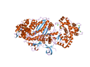
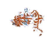
 Generic protein structure example |
Myosin VI, also known as MYO6, is a protein. It has been found in humans, mice, fruit flies (Drosophila melanogaster), and nematodes (Caenorhabditis elegans).
Function
Myosin VI is a molecular motor involved in intracellular vesicle and organelle transport. It is one of the so-called unconventional myosins.[supplied by OMIM][1]
Interactions
MYO6 has been shown to interact with GIPC1[2][3], DAB2.[4][5], ubiquitin[6], and clathrin[7]
References
- ↑ "Entrez Gene: MYO6 myosin VI". https://www.ncbi.nlm.nih.gov/sites/entrez?Db=gene&Cmd=ShowDetailView&TermToSearch=4646.
- ↑ "Large-scale mapping of human protein-protein interactions by mass spectrometry". Molecular Systems Biology 3 (1): 89. 2007. doi:10.1038/msb4100134. PMID 17353931.
- ↑ "Myo6 facilitates the translocation of endocytic vesicles from cell peripheries". Molecular Biology of the Cell 14 (7): 2728–43. Jul 2003. doi:10.1091/mbc.E02-11-0767. PMID 12857860.
- ↑ "Myosin VI binds to and localises with Dab2, potentially linking receptor-mediated endocytosis and the actin cytoskeleton". Traffic 3 (5): 331–41. May 2002. doi:10.1034/j.1600-0854.2002.30503.x. PMID 11967127.
- ↑ "DOC-2/DAB2 is the binding partner of myosin VI". Biochemical and Biophysical Research Communications 292 (2): 300–7. Mar 2002. doi:10.1006/bbrc.2002.6636. PMID 11906161.
- ↑ "Myosin VI Contains a Compact Structural Motif that Binds to Ubiquitin Chains.". Cell Reports 14 (11): 2683–94. 2016. doi:10.1016/j.celrep.2016.01.079. PMID 26971995.
- ↑ "Clathrin light chain A drives selective myosin VI recruitment to clathrin-coated pits under membrane tension.". Nature Communications 10 (1): 4974. 2019. doi:10.1038/s41467-019-12855-6. PMID 31672988.
Further reading
- "Myosin VI, a new force in clathrin mediated endocytosis". FEBS Letters 508 (3): 295–9. Nov 2001. doi:10.1016/S0014-5793(01)03065-4. PMID 11728438.
- "Myosin VI: two distinct roles in endocytosis". Journal of Cell Science 116 (Pt 17): 3453–61. Sep 2003. doi:10.1242/jcs.00669. PMID 12893809.
- "The mouse Snell's waltzer deafness gene encodes an unconventional myosin required for structural integrity of inner ear hair cells". Nature Genetics 11 (4): 369–75. Dec 1995. doi:10.1038/ng1295-369. PMID 7493015.
- "Identification and overlapping expression of multiple unconventional myosin genes in vertebrate cell types". Proceedings of the National Academy of Sciences of the United States of America 91 (14): 6549–53. Jul 1994. doi:10.1073/pnas.91.14.6549. PMID 8022818.
- "Prediction of the coding sequences of unidentified human genes. VII. The complete sequences of 100 new cDNA clones from brain which can code for large proteins in vitro". DNA Research 4 (2): 141–50. Apr 1997. doi:10.1093/dnares/4.2.141. PMID 9205841.
- "Characterization of unconventional MYO6, the human homologue of the gene responsible for deafness in Snell's waltzer mice". Human Molecular Genetics 6 (8): 1225–31. Aug 1997. doi:10.1093/hmg/6.8.1225. PMID 9259267.
- "The localization of myosin VI at the golgi complex and leading edge of fibroblasts and its phosphorylation and recruitment into membrane ruffles of A431 cells after growth factor stimulation". The Journal of Cell Biology 143 (6): 1535–45. Dec 1998. doi:10.1083/jcb.143.6.1535. PMID 9852149.
- "Myosin VI is an actin-based motor that moves backwards". Nature 401 (6752): 505–8. Sep 1999. doi:10.1038/46835. PMID 10519557.
- "Myosin VI isoform localized to clathrin-coated vesicles with a role in clathrin-mediated endocytosis". The EMBO Journal 20 (14): 3676–84. Jul 2001. doi:10.1093/emboj/20.14.3676. PMID 11447109.
- "MYO6, the human homologue of the gene responsible for deafness in Snell's waltzer mice, is mutated in autosomal dominant nonsyndromic hearing loss". American Journal of Human Genetics 69 (3): 635–40. Sep 2001. doi:10.1086/323156. PMID 11468689.
- "Dual regulation of mammalian myosin VI motor function". The Journal of Biological Chemistry 276 (43): 39600–7. Oct 2001. doi:10.1074/jbc.M105080200. PMID 11517222.
- "Myosin VI is a processive motor with a large step size". Proceedings of the National Academy of Sciences of the United States of America 98 (24): 13655–9. Nov 2001. doi:10.1073/pnas.191512398. PMID 11707568.
- "DOC-2/DAB2 is the binding partner of myosin VI". Biochemical and Biophysical Research Communications 292 (2): 300–7. Mar 2002. doi:10.1006/bbrc.2002.6636. PMID 11906161.
- "Myosin VI binds to and localises with Dab2, potentially linking receptor-mediated endocytosis and the actin cytoskeleton". Traffic 3 (5): 331–41. May 2002. doi:10.1034/j.1600-0854.2002.30503.x. PMID 11967127.
- "Interaction of SAP97 with minus-end-directed actin motor myosin VI. Implications for AMPA receptor trafficking". The Journal of Biological Chemistry 277 (34): 30928–34. Aug 2002. doi:10.1074/jbc.M203735200. PMID 12050163.
- "Mutations of MYO6 are associated with recessive deafness, DFNB37". American Journal of Human Genetics 72 (5): 1315–22. May 2003. doi:10.1086/375122. PMID 12687499.
- "Myo6 facilitates the translocation of endocytic vesicles from cell peripheries". Molecular Biology of the Cell 14 (7): 2728–43. Jul 2003. doi:10.1091/mbc.E02-11-0767. PMID 12857860.
External links
- Overview of all the structural information available in the PDB for UniProt: Q9UM54 (Unconventional myosin-VI) at the PDBe-KB.


